
The underwater world is full of wonders, but few are as mind-blowing as regeneration—the ability to regrow lost or damaged body parts. While humans can only scar over wounds, many marine animals (and a few fully aquatic ones) rebuild limbs, organs, or even entire bodies with astonishing precision and speed. This superpower often serves as defense (autotomy: deliberately shedding parts to escape predators) or recovery from injury.
Here are 10 remarkable animals that can regrow body parts underwater, showcasing nature’s incredible repair toolkit. These examples highlight echinoderms, cephalopods, and other marine masters of regeneration.
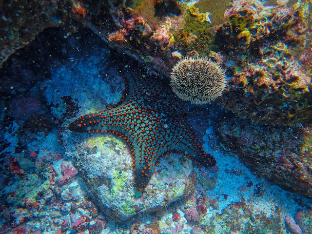
1. Sea Stars (Starfish)
Sea stars are regeneration superstars. They can regrow entire arms lost to predators or accidents, often in months. Some species (like certain Asterias or Linckia) go further: if an arm breaks off with a portion of the central disc (containing vital organs), that arm can regenerate a whole new sea star! Most organs are distributed in the arms, making this possible. This ability helps them survive attacks from birds, fish, or humans.
2. Axolotl (Mexican Walking Fish)
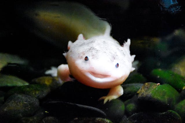
Though technically an amphibian native to Mexican lakes (permanently aquatic due to neoteny—retaining juvenile features like gills), the axolotl is a freshwater icon of regeneration. It regrows entire limbs, tails, spinal cord sections, heart tissue, jaws, eyes, and even parts of the brain—scar-free and repeatedly throughout life. A lost leg can fully reform in weeks, complete with bones, muscles, nerves, and blood vessels. Scientists study axolotls extensively for insights into human tissue repair.
3. Octopuses
Octopuses regrow lost arms (tentacles) with full functionality, including suckers, nerves, and sensory capabilities. The process is complex: a blastema (mass of undifferentiated cells) forms at the stump, rebuilding the arm over weeks to months. Regenerated arms regain full strength and dexterity for hunting and manipulation. This helps them escape predators or survive fights.
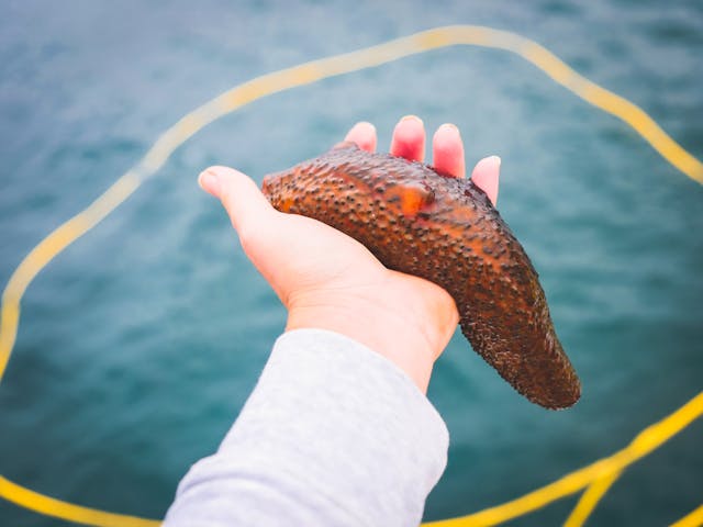
4. Sea Cucumbers
Sea cucumbers eject internal organs (evisceration) as a defense against predators—literally “throwing up” their guts to distract attackers—then regrow them in as little as a week to several weeks. They regenerate the entire digestive system, respiratory trees, and other viscera from the remaining body wall. Some species heal deep wounds rapidly too.
5. Brittle Stars
Close relatives of sea stars, brittle stars (ophiuroids) shed arms easily (autotomy) when threatened. They regrow arms quickly—often faster than sea stars—complete with tube feet for movement and feeding. Some can regenerate multiple arms simultaneously.
6. Crabs and Lobsters
Many crustaceans, including crabs and lobsters, regrow lost legs, claws, antennae, and even eyes through molting cycles. The new appendage starts small but grows larger with each molt until full size. This helps them escape predators that grab limbs.
7. Sea Anemones
Some sea anemones regenerate lost body parts or even entire individuals from fragments. They can regrow tentacles, oral discs, or sections after injury or fission. Certain species show whole-body regeneration from small pieces.
8. Sea Slugs (Certain Sacoglossans)
Species like Elysia atroviridis can deliberately detach their heads (including brain) and regrow an entire new body from the head downward—sometimes multiple times! The head survives on stored nutrients or photosynthesis (from algal symbionts) while regenerating the torso, heart, and organs.
9. Zebrafish (in Freshwater, but Aquatic)
Zebrafish regenerate fins, spinal cord sections, heart muscle, retina, and parts of the brain—making them lab favorites for studying regeneration. Though not strictly marine, their aquatic prowess inspires marine research parallels.
10. Marine Worms (e.g., Platynereis dumerilii)
Certain polychaete worms like Platynereis regrow posterior body segments, including nerves and organs, after amputation. Recent studies (2024) show they reprogram differentiated cells into stem-like states for rapid rebuilding—without relying on pre-existing stem cells.
These animals use mechanisms like blastema formation (a proliferative cell mass), stem cell activation, dedifferentiation (cells revert to a stem-like state), and precise gene networks—far beyond human scarring. Research into these processes could one day help treat human injuries, spinal damage, or organ failure.
The ocean’s “immortals under the knife” remind us how evolution favors survival through renewal. Which of these regenerating marvels fascinates you most? Have you spotted any (like a regenerating starfish) while diving?
Blastema formation is a key biological process in regeneration, where a mass of proliferative, relatively undifferentiated cells (progenitor cells) accumulates at the site of injury or amputation. This structure, called the blastema, acts as a temporary “bud” or outgrowth that proliferates, patterns, and differentiates to rebuild lost tissues or entire body parts—such as limbs, fins, arms, or organs—often with remarkable fidelity.
The blastema is one of the hallmarks of true regeneration (vs. simple wound healing with scarring), seen in highly regenerative animals like axolotls, sea stars, planarians, zebrafish, and some insects. In contrast, mammals (including humans) rarely form a functional blastema after major injury, leading to scarring instead.
General Steps in Blastema Formation
Blastema formation follows a coordinated sequence of events triggered by injury:
- Wound Healing and Closure Immediately after amputation or injury, epidermal cells migrate rapidly to cover the wound, forming a wound epidermis (WE). This seals the site and prevents infection while creating a specialized signaling center.
- Apical Epithelial Cap (AEC) Formation In many models (e.g., axolotls), the wound epidermis thickens and reorganizes into a specialized structure called the apical epithelial cap (AEC). The AEC is crucial—it secretes growth factors and maintains an environment that promotes dedifferentiation and proliferation beneath it. Nerve signaling and factors like FGFs (fibroblast growth factors) and BMPs (bone morphogenetic proteins) are often required for AEC maturation.
- Dedifferentiation and Progenitor Cell Recruitment Mature cells near the injury site undergo dedifferentiation—reverting to a more stem-like or progenitor state (e.g., losing specialized features like muscle striations or becoming multipotent). This includes:
- Histolysis (breakdown of tissues to release cells).
- Activation of resident stem/progenitor cells.
- Migration of cells from surrounding tissues toward the wound.
- Blastema Accumulation and Growth Dedifferentiated or progenitor cells proliferate massively under the AEC, forming a visible, cone- or bud-shaped mass (the blastema). This phase involves:
- High cell division rates.
- Signaling pathways like Wnt, FGF, ERK, and others to drive proliferation.
- Establishment of positional information (proximal-distal, anterior-posterior axes) for correct patterning.
- Redifferentiation and Patterning Once formed and grown, the blastema transitions to redifferentiation: cells re-specialize into muscle, bone, nerves, skin, etc., following developmental-like cues to rebuild the missing structure.
The entire process can take weeks to months, depending on the species and appendage size.
Examples in Specific Animals
- Axolotl Limb Regeneration (classic model): After amputation, wound epidermis forms within hours → AEC develops (innervated) → dedifferentiation of stump tissues (muscle, connective, etc.) → blastema cells aggregate and proliferate beneath the AEC → cone-shaped blastema grows → patterning and redifferentiation rebuild the limb (including bones, muscles, nerves). Lineage-restricted progenitors (not fully pluripotent) contribute, with nerves essential for blastema initiation.
- Sea Stars (Starfish) Arm Regeneration: Regeneration is often more morphallactic (reorganization of existing tissues) than blastema-driven. A “blastema-like” region forms with undifferentiated cells, ECM remodeling, and cell influx from coelom, but it’s less localized and organized than in salamanders. No true blastema in some species; regeneration relies heavily on dedifferentiation and migration.
- Other Cases: In zebrafish fins or planarian heads/tails, similar progenitor accumulation occurs, often involving ERK/CK-2 pathways or wound epidermis signals.
Molecular and Genetic Insights
- Key pathways: FGF, Wnt, BMP, ERK signaling for proliferation; nerves provide trophic factors.
- Epigenetic changes allow dedifferentiation.
- Recent studies (e.g., 2024 cockroach/zebrafish) highlight conserved ERK-activated CK-2 for triggering blastema via DNA replication and cell cycle entry.
In short, blastema formation transforms a wound into a regenerative organ by recruiting and reprogramming cells into a proliferative, patterned mass—nature’s version of a “reset and rebuild” button. This process inspires biomedical research for human regenerative medicine (e.g., scarless healing or limb regrowth).Human Regenerative Medicine in 2026: From Lab Breakthroughs to Clinical Reality
Regenerative medicine aims to repair, replace, or regenerate damaged tissues and organs, shifting medicine from symptom management to true restoration. By harnessing stem cells, gene editing, tissue engineering, and advanced biomaterials, it targets chronic diseases, injuries, aging-related decline, and organ failure. In 2026, the field has transitioned from promising research to scalable, evidence-based therapies, with growing clinical adoption, regulatory progress, and massive market momentum.
Current Market and Growth Snapshot
The global regenerative medicine market has exploded:
- Valued at around $88–102 billion in 2025–2026.
- Projected to reach $340 billion by 2035 (CAGR ~14–17%).
- Key drivers: rising chronic diseases, organ shortages, stem cell/gene therapy advances, and investments in manufacturing infrastructure.
Major players (e.g., Bayer, Sumitomo Pharma, Novo Nordisk) pour hundreds of millions into scalable production facilities for cell therapies.
Key Advances and Milestones in 2026
- Stem Cell Therapies “Come of Age” Japan leads with conditional marketing approvals for the world’s first induced pluripotent stem cell (iPSC)-derived treatments—e.g., for Parkinson’s (Sumitomo Pharma) and cardiac repair (Cuorips). These use patient-derived or allogeneic iPSCs to generate functional cells for transplantation. Over 1,200 patients have received human pluripotent stem cell products in trials by late 2024–2025, with no major safety signals like teratoma formation in long-term follow-up.
- FDA and Regulatory Shifts The U.S. FDA approves more cell therapies (at least two predicted in 2026), some with limited Phase 3 data under accelerated pathways. RMAT (Regenerative Medicine Advanced Therapy) designations speed trials for conditions like lupus nephritis (e.g., iPSC-derived CAR-T cells). The NIH pauses new human embryonic stem cell line approvals, seeking input on alternatives like iPSCs to reduce reliance on embryos.
- Organoids and Lab-Grown Tissues Vascularized organoids (mini-organs with blood vessels) from human pluripotent stem cells advance for heart, liver, and other models. These enable better drug testing, disease modeling, and potential transplantation of engineered patches. Early clinical trials test lab-grown liver/kidney tissue and cardiac patches.
- In-Utero and Specialized Therapies World’s first in-utero stem cell therapy for fetal spina bifida (CuRe Trial, UC Davis) proves safe in Phase 1—combining fetal surgery with placenta-derived stem cells to repair spinal defects before birth.
- Broader Applications
- MSC therapies combat inflammation in age-related diseases (heart, neurodegeneration).
- Gene-edited cells target metabolic disorders, eye diseases (e.g., macular degeneration), and autoimmune conditions.
- Neural stem cell trials for Huntington’s disease launch mid-2026.
- Exosome and cell-free approaches emerge for regeneration without full cell transplants.
- Infrastructure and “Bio-Liquidity” Concepts like preserving younger stem cells (“bio-liquidity”) gain traction for lifelong regenerative use. Automated manufacturing (e.g., hospital-based iPSC foundries) and investments make personalized therapies more accessible.
Challenges and Realistic Outlook
While exciting, regenerative medicine isn’t a cure-all yet:
- Many therapies remain in early trials or conditional approval (safety long-term data still accumulating).
- Immune rejection, scalability, and high costs persist.
- Ethical/regulatory debates continue (e.g., embryonic vs. iPSC sources).
- Unproven clinics market hype—patients should stick to FDA/approved trials.
Success rates vary: 70–92% in approved areas (e.g., joint/cartilage repair, certain cancers via CAR-T), but experimental uses demand caution.
The Future Horizon
2026 marks a pivot: regenerative medicine evolves from “breakthrough science” to “system-ready growth.” With converging biology, regulation, and infrastructure, it promises healthier aging, reduced organ waitlists, and personalized fixes for degenerative conditions.
In short, we’re entering an era where medicine rebuilds what disease or injury destroys—starting with stem cells, organoids, and engineered tissues. The ocean-inspired regeneration we discussed earlier (e.g., axolotls, sea stars) inspires this human quest.
What aspect intrigues you most—stem cell approvals, organoids, or potential for aging reversal? Or want details on a specific therapy?
Induced Pluripotent Stem Cell (iPSC) Therapies: Current Status and Advancements (as of March 2026)
Induced pluripotent stem cells (iPSCs) are adult cells (typically skin or blood) reprogrammed back to an embryonic-like pluripotent state using factors like Oct4, Sox2, Klf4, and c-Myc (the Yamanaka factors, discovered in 2006). This breakthrough allows iPSCs to differentiate into virtually any cell type, making them a cornerstone of regenerative medicine. They enable patient-specific (autologous) or off-the-shelf (allogeneic) therapies, disease modeling, drug screening, and tissue repair without ethical issues tied to embryonic stem cells.
In 2026, iPSC therapies have reached a historic milestone: moving from experimental trials to regulated clinical use, particularly in Japan, while global pipelines expand rapidly.
Landmark Approvals: The World’s First iPSC-Derived Therapies
In February 2026, Japan’s Ministry of Health, Labour and Welfare (MHLW) expert panel recommended conditional and time-limited marketing approval (under the conditional approval pathway for regenerative medicines) for two allogeneic iPSC-derived products—the first such approvals globally. These are expected to be formally authorized soon, with post-market surveillance required for up to 7 years to confirm long-term efficacy and safety.
- Amchepry (raguneprocel) — Developed by Sumitomo Pharma Co. in collaboration with Racthera Inc. (and rooted in research from Kyoto University’s Center for iPS Cell Research and Application, CiRA). This involves transplanting iPSC-derived dopaminergic neural progenitor cells into the brain to replace lost dopamine-producing neurons in Parkinson’s disease. Early trials (e.g., led by Jun Takahashi) showed safety and functional improvements in patients after 24 months, with no major adverse events like tumor formation.
- ReHeart (also referred to as RiHEART or IPSOC-1) — Developed by Cuorips Inc. (a spinout from Osaka University research led by Yoshiki Sawa). This is an ultra-thin sheet (patch) of iPSC-derived cardiomyocytes transplanted onto the heart surface to treat severe heart failure from ischemic cardiomyopathy. It promotes angiogenesis, tissue repair, and improved cardiac function. Tested in small cohorts (e.g., 8 patients), it has shown encouraging results without serious complications.
These approvals mark the transition of iPSC technology from academic promise to commercial regenerative medicine, leveraging Japan’s accelerated regulatory framework for cell therapies.
Global Clinical Trial Landscape
As of early 2026, dozens of iPSC-based trials are active or completed worldwide (estimates: 10+ published studies and 20–30+ ongoing registered trials as of late 2025 data). Applications span:
- Neurological — Parkinson’s (e.g., USC Keck Medicine early-phase trial implanting iPSC-derived dopamine neurons; Keio University trials for spinal cord injury).
- Ocular — Retinal pigment epithelium (RPE) implants for geographic atrophy/AMD (NIH/NEI Phase I/IIa trial); retinitis pigmentosa.
- Cardiovascular — Cardiomyocyte patches/sheets for heart failure.
- Immuno-Oncology — iPSC-derived NK cells and CAR-NK/CAR-T therapies (e.g., Century Therapeutics’ CNTY-101 for lupus; Fate Therapeutics’ FT819/FT522/others for cancers and autoimmune diseases; multiple candidates like CTH-401, NCR300 in pipelines from 12+ companies).
- Other — Platelets for transfusion, graft-versus-host disease, metabolic disorders, and more.
Key players include Fate Therapeutics (multiple off-the-shelf iPSC immune cell products), Century Therapeutics, BlueRock Therapeutics (Bayer), Cynata Therapeutics (iPSC-MSCs for osteoarthritis in Phase 3), and emerging firms like Cellistic (new GMP platforms for iPSC T cells, cardiomyocytes, endothelial cells).
Safety profile remains encouraging: No widespread teratoma or tumor risks in long-term follow-ups from early trials. Challenges include immune rejection (mitigated by HLA-matching or gene editing), scalability, manufacturing costs, and ensuring functional integration.
Advancements and Future Outlook
- Manufacturing — Automated GMP platforms (e.g., CiRA Foundation’s FiT facility, Cellistic’s Echo series) improve reproducibility and scale.
- Conferences & Collaborations — The ISSCR-JSRM Joint Collaboration on Clinical iPSCs launched in 2026; iPSC 20th Anniversary Symposium in Kyoto (October 2026) will highlight progress.
- Broader Impact — iPSCs enable personalized medicine, reduce animal testing via organoids, and inspire in vivo reprogramming approaches.
While most therapies remain in Phase I/II (small, often uncontrolled), the Japanese approvals signal maturation. Expect more data readouts, expanded indications, and potential U.S./EU pathways in the coming years.
iPSC therapies represent a paradigm shift toward repairing rather than managing disease—directly inspired by regeneration in animals like axolotls. What specific condition or trial interests you most (e.g., Parkinson’s, heart failure, or cancer applications)?


Leave a Reply